Falloposcope cancer screening tool advances to next steps
Jennifer Barton spearheads effort to diagnose ovarian cancer earlier and expand treatment options.

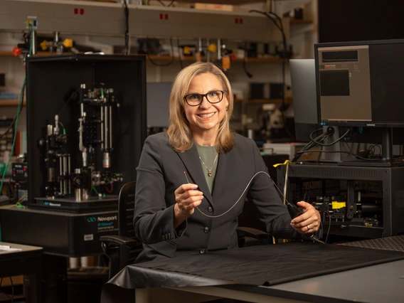
Jennifer Barton, who holds the Thomas R. Brown Distinguished Chair of Biomedical Engineering, has 20 years of experience developing tiny imaging devices that see inside the body.
Kris Hanning, U of A Office of Research and Partnerships
Jennifer Barton, who holds the Thomas R. Brown Distinguished Chair of Biomedical Engineering, has spent decades working to improve early detection of ovarian cancer. Now, she’s advanced to two pilot trials with optimized versions of her patented falloposcope – the first endoscope to image and collect cells from fallopian tubes.
The device will be tested on 200 patients, building on previous data from about 20 participants. Barton and her team plan to refine the device and move it toward clinical adoption.
Helping women with cancer
The falloposcope journey began 20 years ago when a gynecological surgeon friend urged Barton, who also serves as interim vice provost for health programs at the University of Arizona, to work on ovarian cancer.
About 12 years ago, the medical community reached a consensus that the deadliest forms of the disease would start in the fallopian tubes. Physicians lacked a way to examine the fallopian tubes at high resolution – they often identified the cancer when it was too late.
The five-year survival rate for American women diagnosed with ovarian cancer before it metastasizes is 92%, but these patients make up only about a quarter of cases, according to the National Cancer Institute. For those whose cancer has spread, the five-year survival rate is 32%.
Barton, also a professor of optical sciences and a member of the BIO5 Institute, invented the falloposcope to save lives with early detection and help high-risk patients avoid unnecessary removal of the ovaries and fallopian tubes.
Genetic testing and family history analysis identify high-risk patients. One common finding is a mutation in BRCA1 or BRCA2 genes, which prevents the genes from repairing damaged DNA.
Currently, women who are at high risk for ovarian cancer have two choices, Barton said.
They can undergo surgery at age 35 or opt for surveillance with blood tests and an ultrasound. But research indicates those methods aren’t reliable for early detection, she added.
“What it comes down to is, you either get the surgery or you take your chances.”
Taking on the challenge
No early detection method exists due to inherent obstacles to reaching the fallopian tubes.
“A key focus area is learning how to thread the endoscope through all of the twists and bends in the fallopian tube,” said Dr. John Heusinkveld, Barton’s longtime clinical research partner and a Banner Health physician and surgeon.
Not only are the tubes twisty and tiny, but they’re also filled with finger-like projections called cilia, which move eggs and sperm. The instrument must have a diameter of less than 1 millimeter and be slick enough to move without catching on cilia.
“It’s a challenge, but it’s absolutely possible with modern technology,” Barton said.
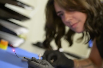
Dilara Long, biomedical engineering doctoral student and MD/PhD candidate at the U of A College of Medicine – Tucson, assists with the diagnostic use of the falloposcope and is exploring additional applications for the device.
She secured initial funding from the U.S. Department of Defense Ovarian Cancer Research Program in 2018. Since then, she has raised additional support from the DOD, the National Cancer Institute and private donors. The team began the first human trial in 2021.
Barton and her team have published studies showing the device is safe and renders high-resolution structural and biochemical images. They’ve also documented the baseline for normal fallopian tube tissue and made improvements based on the clinical experiences of Heusinkveld, also an associate professor of obstetrics and gynecology at the College of Medicine – Tucson.
One trial, conducted locally in partnership with Banner Health, focuses on 10 participants at normal risk who are having surgery for reasons other than cancer. The other trial is underway at New York-Presbyterian Queens Hospital, which runs a clinic for high-risk patients. Collaborators there are using the falloposcope to test tissue directly after its removal in surgery.
The path to patients
Barton estimated current trials will conclude in 2027. Next, she will find an industry partner to license the invention, conduct additional studies and obtain Food and Drug Administration approval.
Barton and Heusinkveld envision falloposcope testing as an outpatient procedure for high-risk patients.
"I lost a good friend to ovarian cancer at age 35. Her mother died young of ovarian cancer, too," Barton said. "I'm quite sure she had a genetic abnormality, and she could have been somebody that maybe we could have helped."
Barton expects the falloposcope will serve additional purposes beyond cancer, including determining causes of infertility like endometriosis.
"I'm proud to work on those devices and techniques that will solve problems," Barton said.“I’m proud to work on those new devices and techniques that will solve problems,” Barton said.