Dongkyun Kang wins $2.4M grant to develop noninvasive microscope
The associate professor of biomedical engineering is working to improve the imaging of cancer patients' nerve endings at the University of Arizona Cancer Center.
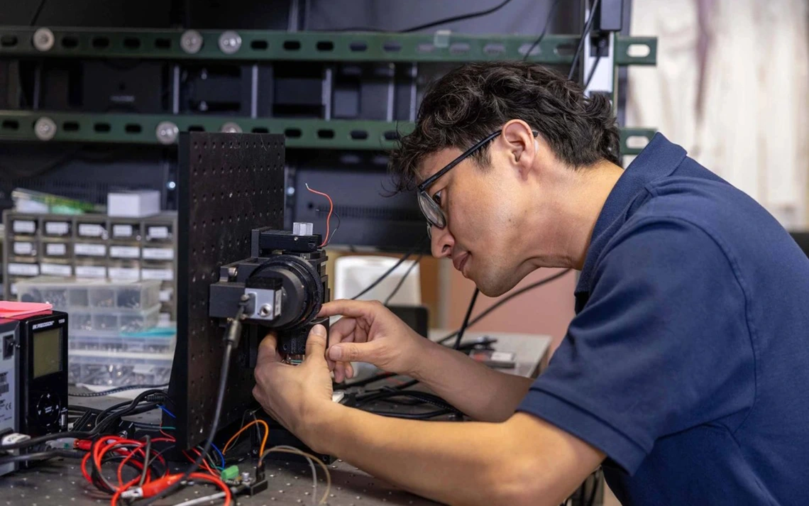
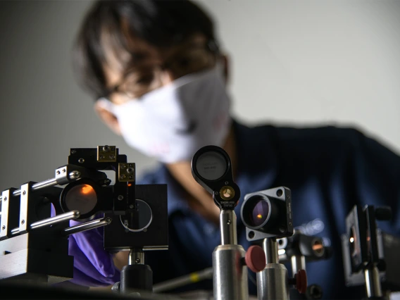
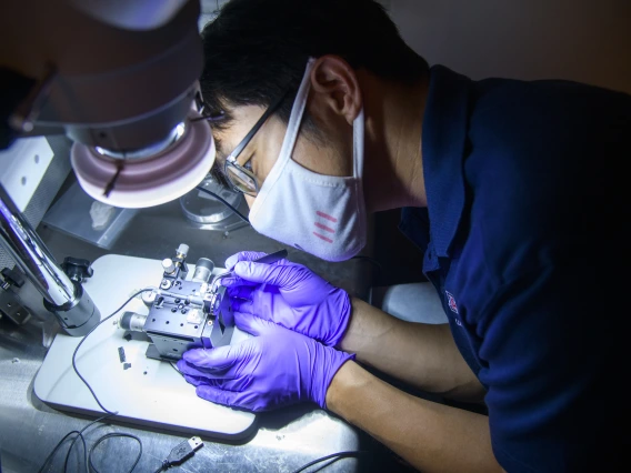
Dongkyun Kang is creating a confocal microscope that may help doctors diagnose chemotherapy-induced peripheral neuropathy earlier, leading to better treatment and prevention opportunities.
Joshua Elz, University of Arizona Cancer Center
The National Cancer Institute granted Dongkyun Kang, an associate professor in the College of Engineering’s Department of Biomedical Engineering, $2.4 million to develop a noninvasive, confocal microscope to examine nerve endings of cancer patients with chemotherapy-induced peripheral neuropathy and identify potential biomarkers for the disease.
Peripheral neuropathy is a common side effect of certain chemotherapy drugs. Chemotherapy-induced peripheral neuropathy, or CIPN, can be severe and debilitating, often causing physical limitations, including numbness, weakness and pain in the hands and feet of patients, and reduced quality of life.
“CIPN symptoms can cause high levels of discomfort and present multiple challenges in the daily lives of cancer patients,” said Kang, who has an additional appointment in the U of A James C. Wyant College of Optical Sciences and is a member of the BIO5 Institute. “Using this approach, we may be able to identify CIPN earlier to stop symptoms from progressing and possibly prevent the condition altogether.”
Patients with CIPN are known to have a reduced number of Meissner corpuscles, which are nerve endings responsible for transmitting the sensations of light touch and low vibration. The confocal microscopy images will be used to find and count Meissner corpuscles, eventually leading to a potential imaging biomarker.
Noninvasive, highly accessible
Kang’s lab has pioneered low-cost confocal microscopy over the past seven years. His team was the first to demonstrate that noninvasive microscopy can be built at low cost, which makes it highly accessible in a wide variety of clinical settings.
“This study will build the evidence that our noninvasive microscopy approach can provide quantitative imaging biomarkers for CIPN monitoring, treatment and research,” Kang said.
Kang said his goal is to change the diagnostic focus from subjective, qualitative markers, like patient and clinician questionnaires, to objective, quantitative biomarkers that could support personalized care for patients with CIPN.
“The work Dr. Kang has been doing exemplifies the Cancer Center’s approach to precision prevention and therapy,” said Dan Theodorescu, the Nancy C. and Craig M. Berge endowed chair for the director of the Cancer Center. “I’m excited to see how his study evolves, especially given its potential for global impact.”