“The Future is Now” in New Skin Cancer Diagnosis Technology
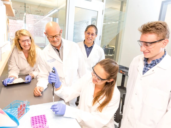
Biomedical engineering professor Dongkyun "DK" Kang is collaborating with fellow researchers in the University of Arizona Cancer Center to create a portable, less expensive version of a skin cancer diagnostic microscope.
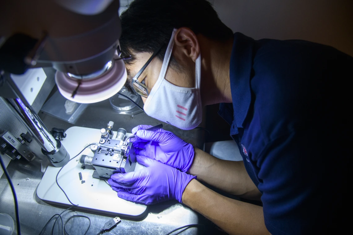
Assistant biomedical engineering professor Dongkyun "DK" Kang has created a portable prototype of a microscope used to help diagnose skin cancer. Here, he works to improve the device assembly.
The sun’s harmful ultraviolet rays pose elevated risks for skin cancer in the desert southwest, but University of Arizona Health Sciences researchers at the Cancer Center are using their unique position to address this disease with new technology.
The Center’s Skin Cancer Institute has a novel device that provides noninvasive imaging of the skin called reflectance confocal microscopy, or RCM. It allows doctors to quickly and safely diagnose many skin cancers and monitor responses to treatment without a biopsy.
Dr. Clara Curiel-Lewandrowski, co-director of the Skin Cancer Institute and interim dermatology division chief at the College of Medicine – Tucson, brought the technology to the university. She is now working with biomedical engineering assistant professor Dongkyun Kang, co-leader of the Cancer Imaging Program, to create a version of the device that is more accessible to doctors trained in skin cancer diagnosis.
The current instrument costs more than $80,000, making it too expensive for many clinical practice settings. This team aims to create a handheld or portable version of the device which costs closer to $1,000.
The technology is made with very sophisticated electrical and optical components, according to Kang, who is also an assistant professor in the James C. Wyant College of Optical Sciences. His lab has been focused on developing a portable confocal microscope, or PCM, using an inexpensive near-infrared LED to lower the manufacturing costs and make the device more practical for clinical use. Kang has also tested a smartphone PCM device at the Infectious Diseases Institute in Uganda.
“The goal is to make it widely available not just to dermatologists, but to practitioners around the world,” Kang said. “Collaboration is very important so that we are making this technology work in the context of the clinical setting. I work very closely with Clara and also my collaborators with the project in Uganda to see what we can improve.”