BIO5 Researchers Make Tools Better and More Accessible
The BIO5 Institute, directed by biomedical engineering professor Jennifer Barton, is the University of Arizona's home for cutting-edge research.
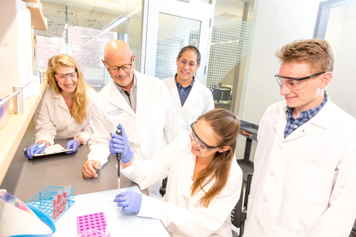
Researchers at BIO5. (Photo: Chris Richards / University of Arizona Alumni Association)
BIO5 connects and mobilizes hundreds of world-class researchers -- including bioscientists specializing in plants, animals and humans as well as engineers, physicians and computational researchers -- to develop creative solutions for complex challenges such as disease, hunger, water and food safety, and other health issues facing Arizona and the world. This interdisciplinary approach has resulted in disease prevention strategies and promising new therapies, innovative diagnostics and devices, and improved food crops.
“We are able to help connect, facilitate and deploy people, resources and funding to produce a result that could help change the course of our community impact related to grand challenges like COVID-19 and other large-scale challenges and crises,” says Jennifer Barton, director of the BIO5 Institute, who works on early cancer detection in her own BIO5 lab.
Barton serves as the chief connector of nearly 350 top scientists from over 70 departments across the university. Their collective bioscience breakthroughs have made possible new technology that is already making a mark in global markets. Barton, who has directed the institute since 2015, works on early cancer detection in her own BIO5 lab. We asked Barton to take us on a conceptual tour of some of BIO5's optics labs.
Medical Imaging and Microfabrication
The tour began with the lab of Tsu-Te "Judith" Su, who works on the microfabrication of nanostructures. “She makes incredibly sensitive detectors of biomarkers in the body. One of her big areas is looking at markers of Alzheimer’s disease.”
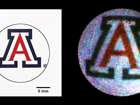
Su’s team is putting photonics technology into a biosensor that could become a useful diagnostic tool. It uses microtoroids, tiny ring-shaped structures, with which a light can be directed to travel around a sample and find very small particles of the protein biomarker being flowed past the sensor.
“She needs to fabricate these devices, and then we need to test them to make sure they are useful as a diagnostic,” Barton says. “Judith works with the physicians and brain scientists here in the BIO5 Institute to make sure her biophotonic device works for this medical problem.”
Optomechanics
The work of Dongkyun "DK" Kang is an ideal example of where optics is going, Barton says. “D.K. is taking the confocal microscope and turning it into a portable handheld device that can go into rural areas — including rural Arizona and into developing countries — to diagnose cancers that affect a lot of people.”
Kang set out to do human tissue imaging in vivo using a smartphone attached to a confocal microscope. That could facilitate easy-to-use, high-resolution imaging in rural clinics at low cost — and ultimately allow for early diagnosis and treatment and lower mortality rates.
He calls it “stopping cancer with a smartphone.” The cancers Kang is targeting include skin cancers, cervical cancers and oral cancers.
“This shows what we are going to do in optomechanics,” Barton says.
The commercial confocal microscope is a large device that can cost upwards of $100,000. Kang is taking that capability and making sensors that are portable and highly sensitive — with a cost goal of $5,000.
“This is all because of advances in photonics,” Barton says. “You can use a small laser diode and implement it in the imaging and diagnostic devices that we are building. Today you can manufacture small optics at a reasonable price.”
Barton predicts an explosion of new technologies coming on the market. “I think we are finally there in terms of products that are truly useful, can truly help patients, and are inexpensive enough to have good market penetration. That’s the difference between the $100,000 device for confocal microscopy and the $5,000 device.”
Translational Imaging
In Barton’s own lab, work on spotting ovarian tumors is unfolding. She is developing what will be the first effective screening method for ovarian cancer. The work, she says, illustrates BIO5’s ability to carry out fabrication and imaging using high resolution optical techniques.
“If you want to look at cells or tissue microstructure, you can’t image very deeply. You end up needing to build endoscopes,” Barton says. “And I do that.”
Her microendoscopes provide nonsurgical techniques for looking into the body’s tissues.
The body has many natural orifices needing study, Barton says, “and the same advances in photonics and materials allow us to make tiny, flexible endoscopes that can go all kinds of places in a body, in a pretty minimally invasive manner.”
Barton’s work, funded by a Department of Defense Ovarian Cancer Research Program grant, has involved placing the devices in the fallopian tubes, where they can do imaging below the surface to a depth of about a millimeter with noninvasive optical coherence tomography, or OCT.
Previous technology only allowed the taking of white light images on the surface. “We are now going to create images like what your eye would see if it could get in there,” Barton says.
The new technology, which Barton says is about seven years away from becoming a market product, will provide the capability to look for cancer below a surface with OCT, to look at cell functions and metabolism with fluorescence, and to look at cell structure with microscopy. “We are adding a great deal of extra capability to what the medical community has had in the past.”